Home
/ Animal Cell Telophase Microscope / File Telophase In Animal Cell Jpg Wikimedia Commons : In plant cells cytokinesis is accomplished by the formation of a cell plate.
Animal Cell Telophase Microscope / File Telophase In Animal Cell Jpg Wikimedia Commons : In plant cells cytokinesis is accomplished by the formation of a cell plate.
Animal Cell Telophase Microscope / File Telophase In Animal Cell Jpg Wikimedia Commons : In plant cells cytokinesis is accomplished by the formation of a cell plate.. Looking at a microscope slide, count and record the number of cells in the field of view that are in each phase. Normal cell division may be observed in onion root tips. Cytokinesis or cytoplasmic division usually occurs at the end of telophase. In eukaryotic (plant, animal & fungus) cells, the division of chromosomes and cytoplasm into two cells is known as the mitotic phase. In plant cells cytokinesis is accomplished by the formation of a cell plate.
If you have a microscope (400x) and a properly stained slide of the onion root tip (or allium root tip), you can see the phases in different cells, frozen in time. Animal cells during mitosis anaphase : Determine the total number of cells counted. The cell on the right is in telophase having nearly completed the segregation of the chromosomes into new daughter cells. Interphase prophase metaphase anaphase telophase & cytokinesis
Quia 9ap Chapter 12 The Cell Cycle Detailed from www.quia.com Mitosis cell in the root tip of onion under a microscope stock. In animal cells, centrioles start. Draw a schematic representation of your observations for both plant and animal cells Look for a cell at telophase. Again switch to low power. Early telophase of mitosis lm stock image p673 0078 science. Mitosis cell in the root tip of onion under a microscope stock. 2be0h26 (rm) mitosis in the onion root tip, telophase, 1000 x optical microscope, photomicrography, genetics.
Normal cell division may be observed in onion root tips.
Animal cell, mitosid model, ultra structure of cell, animal cell division mitosis, three dimensional model, early prophase, prophase, late prophase, metaphase, anaphase, late anaphase, telophase, structure of an animal cell, endoplasmatic reticulum, mitochondria, ribosomes respectively polysomes, golgi apparatus, mitotic cell division, images of animal cell Produkte für gewerbe und wissenschaft. Even though the cells in these tissues are rapidly dividing, most of the cells you see will be in interphase (between cell divisions). Blastula, an animal embryo at the stage immediately following the division of the fertilized e id: In the space below, compare and contrast animal and plant cells during telophase by drawing what you view under the microscope and labeling the following parts: Mikroskop beim führenden marktplatz für gebrauchtmaschinen kaufen. Mitosis cell in the root tip of onion under a microscope stock. Viewing animal cells under a microscope. In this article we will discuss about the stages of mitosis. You will be looking at strands of dna inside the cell! In the telophase stage of mitosis in plant cells, locate the cell plate and explain the purpose and function of the cell plate in your conclusion.) 9. A full set of chromosomes reaches each pole of the cell. Chromosomes nuclear membrane cytoplasm cell plate
Produkte für gewerbe und wissenschaft. Microscope whitefish or fish blastula slide procedure: Blastula, an animal embryo at the stage immediately following the division of the fertilized e id: 2be0h26 (rm) mitosis in the onion root tip, telophase, 1000 x optical microscope, photomicrography, genetics. A full set of chromosomes reaches each pole of the cell.
Mitosis Scientist Cindy from www.scientistcindy.com Cytokinesis or cytoplasmic division usually occurs at the end of telophase. In plant cells cytokinesis is accomplished by the formation of a cell plate. Microscope whitefish or fish blastula slide procedure: Somatic cells make up most of your body's tissues and organs, including skin, muscles, lungs, gut, and hair cells. Mikroskop beim führenden marktplatz für gebrauchtmaschinen kaufen. Mitosis cell in the root tip of onion under a microscope stock. The cell on the right is in telophase having nearly completed the segregation of the chromosomes into new daughter cells. Presented in the digital fluorescence micrograph above is a pair of rat kangaroo (ptk2) kidney epithelial cells in the late stages of telophase.
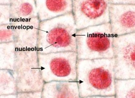
Even though the cells in these tissues are rapidly dividing, most of the cells you see will be in interphase (between cell divisions).
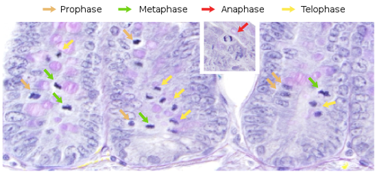
In this article we will discuss about the stages of mitosis. The chromatin is stained with a blue fluorescent probe (dapi), while the cytoskeletal microtubule network and mitotic spindle are stained green (alexa fluor 488). Using a microscope to view the phases of mitosis overview in this exercise you will explore the stages of mitosis using the bionetwork virtual microscope to visualize and identify each stage in both onion root tip and whitefish blastula slides. The nucleus and nucleoli begin to reappear. Where a ring of actin fibers forms around the cell equator and contracts to pinch the cell into two parts. 2be0h26 (rm) mitosis in the onion root tip, telophase, 1000 x optical microscope, photomicrography, genetics. Using your microscope, scan the slides to find a cell in interphase and each one of the four stages of mitosis. The four main phases of mitosis are prophase, metaphase, anaphase, and telophase. Examine prepared microscope slides of both animal cells (whitefish blastula) and plant cells (onion/allium root tip). I brought the microscope into focus on the only the nucleus is replicated during mitosis, and it migrates to the ends of the cell. Animal cells separate by forming a cleavage furrow. Blastula, an animal embryo at the stage immediately following the division of the fertilized e id: Animal mitosis under the microscope.
Animal mitosis under the microscope. Early telophase of mitosis lm stock image p673 0078 science. A brief mitotic phase in which the cell divides its nuclear and cytoplasmic contents, and a longer period between divisions called interphase. Locate cells under low power, then focus the cells at high power. If you have a microscope (400x) and a properly stained slide of the onion root tip (or allium root tip), you can see the phases in different cells, frozen in time.
The Cell 8 Cell Cycle Atlas Of Plant And Animal Histology from mmegias.webs.uvigo.es The cell on the right is in telophase having nearly completed the segregation of the chromosomes into new daughter cells. Cytokinesis is also nearly complete in this cell, with the remnants of the polar microtubules, the midbody, being pinched by the progression of the cytokinetic furrow. Mitosis is the usual form of nuclear division and occurs universally amongst the somatic cells (soma=body) of higher animals. How a cell divides to make two genetically identical cells. Students know the characteristics that distinguish plant cells from animal cells, including chloroplasts and cell walls.: The mitotic spindle begins to disappear. Determine the total number of cells counted. Cytokinesis is the division of the cytoplasm that often begins during late anaphase or early telophase and separates the nuclei to form two daughter cells.
They are both eukaryotic cells, they both have a nucleus, membrane bound organelles, and a cell membrane.
Produkte für gewerbe und wissenschaft. Students know cells divide to increase their numbers through a process of mitosis, which results in two daughter cells with identical sets of chromosomes. Blastula, an animal embryo at the stage immediately following the division of the fertilized e id: Again switch to low power. Because prepared slides are used, these cell divisions have been frozen in time. The mitotic spindle begins to disappear. Examine the cell under high power. Calculate the time (in minutes) for each phase by multiplying the percent of cells in that phase by the number of minutes for the whole cycle. Describe the similarities and differences. Animal cells separate by forming a cleavage furrow. Mikroskop beim führenden marktplatz für gebrauchtmaschinen kaufen. Even though the cells in these tissues are rapidly dividing, of the cells you. Animal mitosis under the microscope.
Looking at a microscope slide, count and record the number of cells in the field of view that are in each phase animal cell telophase. Students know the characteristics that distinguish plant cells from animal cells, including chloroplasts and cell walls.:
Share :
Post a Comment
for "Animal Cell Telophase Microscope / File Telophase In Animal Cell Jpg Wikimedia Commons : In plant cells cytokinesis is accomplished by the formation of a cell plate."


Post a Comment for "Animal Cell Telophase Microscope / File Telophase In Animal Cell Jpg Wikimedia Commons : In plant cells cytokinesis is accomplished by the formation of a cell plate."